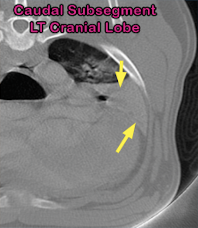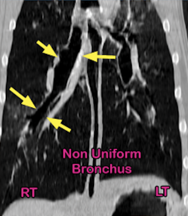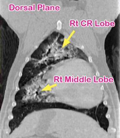
Patsy’s Pulmonary Changes
6-year-old American Bulldog with left-sided, muffled lung sounds received thorax CT scan to determine the cause.

Stay Tuned!
We'll be sharing more case studies soon. Check this page frequently to stay up-to-date on all the excitement happening with MACT!